 Grey matters and nerve bundles within the brain
Are you curious about how your brain works, what its inner structures look like, and how it influences your physical behaviors? Well, Medical Image Illustrator (MiiL), the new medical image processing software developed at the NCHC, will help unravel the mysteries of the human brain by visualizing it in 3D stereo! Using MiiL, researchers will be able to observe the features of the inner brain from an unprecedented view and in extremely high resolution!
 Documentary on Dr. Isaac Tseng
Field Research and Development of Intelligence Drives 3D Medical Image Processing to a New Level!
MiiL is a special medical image processing software developed by the NCHC's Visualization and Interactive Media Laboratory (VIML). MiiL pROVides diverse medical image processing and 3D real-time display. It also supports all popular medical image formats, massive data processing, 4D data processing, and stereoscopic display.
MiiL makes medical imaging research and processing much more convenient, impROVes medical service quality, and greatly reduces the costs associated with medical research. In Taiwan alone, MiiL has saved domestic research institutes USD10,000-15,000 in commercial software costs and USD2,500-3,300 in annual maintenance costs. Additionally, since the MiiL software was developed in Taiwan and is primarily used there, it can be easily customized for special domestic research needs and saves time in that it reduces the waiting time normally associated with commercial software updates.
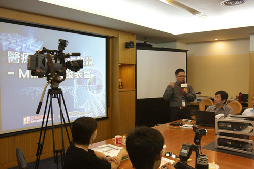 Press interview
MiiL--Taking Brain Research and Clinical Medical Treatment to the Next Level!
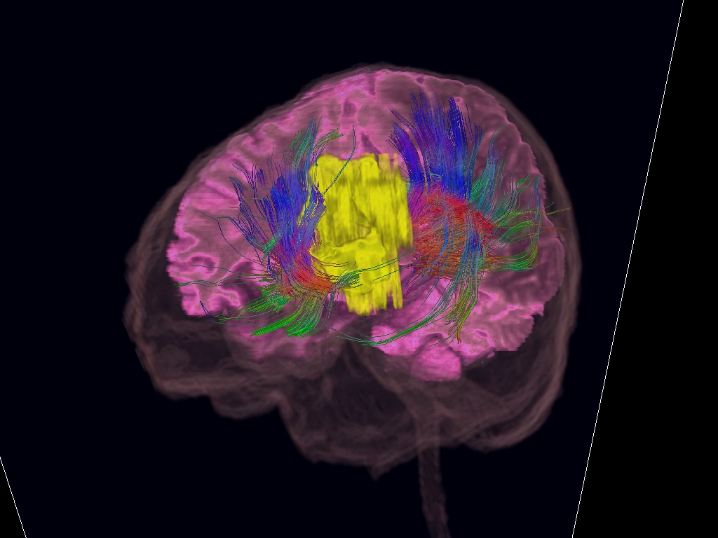
The current cooperation between the NCHC's VIML and the Nuclear Magnetic Resonance Laboratory at National Taiwan University Hospital (a.k.a. the Laboratory) is mutually beneficial. The Laboratory is equipped with the most advanced water molecule diffusion MRI technology in the world in that it is able to capture extremely fine cerebral nerve tract images, however, it cannot integrate with other MRI imaging technologies such as skull, cerebral grey matter, or tumor images, or pROVide real-time 3D display. In order to address these limitations, the Laboratory sought to cooperate with the NCHC's VIML.
The NCHC's VIML used MiiL to visualize various brain structures including the shape of the skull, cerebral grey matter and nerve tract information, tumors. MiiL was also used to integrate all data for calibration of contrapositions. The VIML used this data to create a staggered 3D presentation of the cerebral nerve tract and cerebral grey matter. These visualization achievements allow medical personnel to clearly see the brain structures. They also allow doctors to make faster and more accurate diagnosis and have potential value in pre-operative planning.
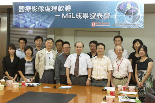 NCHC's MiiL project team
The NCHC to PROVide Download Services for MiiL Medical Imaging Software
In order to satisfy the demand for the MiiL software, the NCHC has created a customized user-friendly interface. The interface offers 2D image reading, marking, and real-time 3D imaging. Very soon, the NCHC will release the MiiL Medical Image processing software, available for all medical researchers to download free of charge.
MiiL supports all popular medical imaging formats. MiiL also greatly reduces the time normally required for image processing which, in tu, allows medical researchers more time for other important R&D. MiiL also supports 3D play equipment, can create stereoscopic images, and will help drive future business opportunities in 3D image equipment manufacturing.